

HPLC MS Analysis for Peptide Research: A 2026 Guide
You're standing at the bench with a peptide vial, a vendor purity claim, and a
Glutathione is unequivocally a tripeptide made of exactly three amino acids: glutamate, cysteine, and glycine. That simple classification matters far more than is commonly understood, because the fact that glutathione is a peptide directly affects how you handle it, measure it, store it, and interpret any experiment that depends on it.
A lot of confusion starts with the question itself. People ask, “Is glutathione a peptide?” as if the answer is just a vocabulary check. In practice, it's a reagent-quality question, a mechanism question, and often a data-integrity question. If you treat glutathione like a generic antioxidant supplement, you'll miss the molecular features that determine whether it survives in solution, whether your assay distinguishes reduced from oxidized material, and whether the biology you observe reflects the cell model or your preparation method.
That's why researchers need to think about glutathione the way they'd think about any critical small biomolecule in a sensitive system. Its chemistry isn't background information. Its chemistry is the experiment.
If you need the direct answer to is Glutathione a peptide, yes. More specifically, it's a tripeptide built from three amino acids linked into a defined small chain.
That answer becomes useful only when you follow the consequences. Peptides aren't just “small protein-like things.” Their bond architecture, side-chain chemistry, and degradation pathways control how they behave in cells and in tubes. Glutathione sits at the center of redox biology, but in the lab it also behaves like a chemically active analyte that can change during preparation, storage, or assay workup.
For a new researcher, the mistake is usually conceptual. They assume that because glutathione is common in biology, it's forgiving in experiments. It isn't always forgiving. If you let oxidation occur during sample handling, your measured redox state may reflect bench exposure rather than cell physiology. If you buy poorly characterized material, you may confuse impurity-driven signal drift with biology. If you select an assay that reads total thiols when your real question is the balance between reduced and oxidized forms, your conclusion may be directionally wrong.
A useful starting point is to understand what researchers mean by research peptides in laboratory use. Glutathione belongs in that conversation because its peptide identity is not incidental. It determines how the molecule survives enzymatic attack, participates in redox reactions, and interacts with analytical methods.
Practical rule: If a molecule's structure determines its redox behavior, then structure, handling, and assay choice must be treated as one integrated problem.
That's the mindset that keeps glutathione work clean. You're not just asking what it is. You're deciding how much of your signal is chemistry, how much is biology, and how much might be artifact.
Why does glutathione behave so differently from many other small peptides you might keep in the same freezer box? The answer starts with its architecture. If you miss that structural detail, it becomes easy to make the wrong call about stability, assay design, or even which material to order.
Glutathione is a tripeptide built from glutamate, cysteine, and glycine. That classification is correct, but it is only the first layer of the answer.
The structural point that matters in practice is the bond between glutamate and cysteine. In most peptides, amino acids connect through the alpha carboxyl group. In glutathione, glutamate links to cysteine through its gamma (γ) carboxyl group. You still have a peptide, but you do not have a standard short peptide with standard geometry.
A more accurate way to picture it is a three-residue molecule assembled with one atypical junction. That altered junction changes how enzymes recognize it, how the molecule persists in biological samples, and how confidently you can borrow assumptions from ordinary peptide workflows.
For a researcher, this is not a naming detail. It is a handling detail.
The γ-peptide bond gives glutathione a different biochemical profile from a typical linear tripeptide. Many common peptidases are tuned to recognize the usual backbone arrangement. Glutathione presents a different structural target, so generic peptide degradation rules do not transfer cleanly.
That is one reason glutathione can remain available in cells as a major small-molecule thiol. In the lab, it also means that losses in signal often come from the wrong place if you are thinking only in peptide terms. Oxidation state, trace metal exposure, buffer composition, pH, freeze-thaw history, and time at room temperature can matter more than broad concern about routine proteolysis.
This distinction affects day-to-day bench work:
Researchers often place glutathione in the wrong mental category. It is small enough to resemble a metabolite and organized enough to qualify as a peptide. In experimental work, you need both views at once. Its peptide structure explains its connectivity and enzyme interactions. Its reactive sulfur and atypical linkage explain why storage, handling, and analytical method choice have such direct consequences for data integrity.
Why does one small tripeptide show up in experiments as different as oxidative stress assays, chemotherapy response studies, and inflammatory models? The answer sits in one atom. Glutathione contains a cysteine thiol, and that sulfur is chemically reactive under physiologic conditions. In the lab, that means glutathione is not just another analyte to quantify. It is part of an active redox circuit that can shift during treatment, extraction, storage, and measurement.
A visual helps anchor the moving parts before you interpret any assay readout.
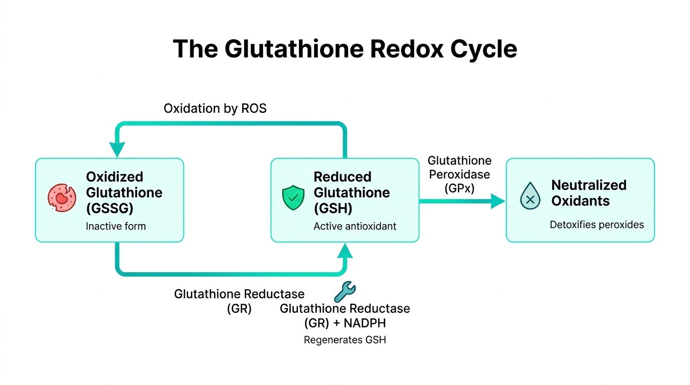
Cells maintain glutathione at relatively high intracellular levels because it is central to day-to-day redox control. Reduced glutathione, or GSH, donates electrons during peroxide detoxification and related reactions, often through glutathione peroxidase. Once oxidized, it can form glutathione disulfide, or GSSG, and cellular reductive systems convert that pool back to GSH.
This cycle matters experimentally because it is fast and condition-sensitive. A delay in quenching, trace metal contamination, repeated freeze-thaw, or a poorly chosen lysis buffer can shift the apparent GSH:GSSG balance before you ever start the assay. If you work on mitochondrial stress, toxicology, inflammatory signaling, or drug response, glutathione can be the readout, part of the mechanism, or a hidden source of variability.
A practical comparison helps here. Glutathione behaves less like a static inventory marker and more like a rechargeable battery that is constantly being discharged and recharged. If you measure it without controlling the handling conditions, you may end up characterizing the sample preparation rather than the biology.
Later in the workflow, this short explainer can help frame the cycle visually.
Outside oncology, glutathione is often discussed as a protective intracellular buffer. In cancer biology, that framing is incomplete. Emerging research shows that many cancer cells, particularly in pancreatic and glioblastoma tumors, overexpress glutathione to neutralize chemotherapy-induced oxidative stress, making glutathione depletion a more effective therapeutic strategy than supplementation, according to the ScienceDirect topic page on glutathione in medicine and oncology.
That changes how you should read the signal. In a treatment-resistant tumor model, higher glutathione can reflect stronger adaptation to therapy-induced stress. Lower glutathione can indicate increased susceptibility to treatment, although it can also arise from broad cytotoxicity or failed metabolic maintenance. The number alone is not enough. You need the disease model, treatment mechanism, sampling time, and compartment being measured.
For bench work, interpretation often goes wrong. Researchers may see an increase in glutathione and label it beneficial, or see a decrease and assume antioxidant failure. In oncology and resistance studies, those shortcuts can invert the biological meaning and push follow-up experiments in the wrong direction.
| Context | High glutathione may suggest | Low glutathione may suggest |
|---|---|---|
| Healthy stressed cell model | stronger redox buffering | oxidative burden or impaired defense |
| Tumor resistance model | survival advantage under therapy | potential therapeutic vulnerability |
| Drug screening assay | target adaptation or compensation | on-target depletion or nonspecific toxicity |
The practical lesson is simple. Glutathione readouts are only as useful as the experimental context around them. If procurement, handling, derivatization, or storage conditions blur the distinction between GSH and GSSG, disease interpretation gets blurred too. That is why the chemistry of this peptide has direct consequences for assay design and data integrity, especially in disease-focused research.
Glutathione is not just supplied to cells from outside. It is synthesized de novo in the liver and found in high concentrations in the cytosol and mitochondria of aerobic organisms, with the chemical formula γ-L-Glu-L-Cys-Gly, as summarized in the Cymbiotika article describing glutathione's composition and distribution.
This changes how you should think about perturbation experiments. If you add glutathione extracellularly and expect a simple intracellular increase, you may be imposing a model that doesn't match the cell's actual regulatory logic. Cells control glutathione through synthesis, utilization, oxidation, regeneration, compartmentalization, and degradation. External addition is only one possible influence on that system.
In practice, intracellular glutathione levels often reflect pathway state more than media supplementation. Substrate availability, oxidative demand, and enzyme activity all matter. When a treatment changes glutathione abundance, the result may come from altered synthesis, altered consumption, altered export, or altered regeneration.
Researchers usually get better data when they design around the glutathione lifecycle rather than a single endpoint. That means asking where the signal originates.
Consider these design questions before starting:
If your intervention changes glutathione, don't assume uptake. Ask whether the cell made less, used more, regenerated less efficiently, or redistributed the pool.
Degradation matters too, even if many studies focus mainly on biosynthesis. Once you adopt a lifecycle view, odd results become easier to troubleshoot. A flat glutathione level after a strong perturbation doesn't always mean “nothing happened.” It may mean synthesis and consumption shifted in opposite directions and temporarily masked each other.
Method choice determines what “glutathione level” means in your data. That phrase can refer to total thiols, reduced glutathione, oxidized glutathione, or a ratio inferred after derivatization and calculation. Those are not interchangeable measurements.
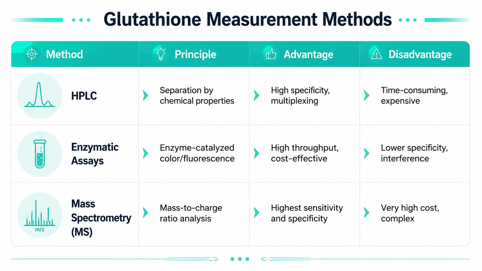
A practical way to choose is to start with the biological question, not the instrument you already have access to.
| Method | Best use case | Main caution |
|---|---|---|
| Ellman-type or total thiol readouts | quick screening when broad sulfhydryl content is sufficient | may not isolate glutathione specifically |
| Glutathione reductase cycling assays | comparing reduced and oxidized pools across many samples | sample preparation strongly affects apparent ratios |
| HPLC | higher-specificity separation when you need clearer analyte identity | more method development and longer runs |
| LC-MS or LC-MS/MS | highest-confidence identification in complex matrices | expensive platform, stricter workflow discipline |
If your question is “Did oxidative stress broadly increase thiol burden changes?” a simple enzymatic or colorimetric workflow may be enough. If your question is “Did this treatment specifically alter GSH versus GSSG in a mixed biological matrix?” then separation-based methods become much more attractive.
For researchers building reliable analytical workflows, this overview of peptide impurity profiling methods in 2026 is useful because glutathione work faces the same underlying problem. Signal quality depends on knowing what else is present, what co-elutes, and what your assay can or cannot distinguish.
Most glutathione assay failures don't come from a bad reader or a bad detector. They come from sample history.
Common problems include:
A good assay answer is always conditional. It means, “This is what the sample contained after this exact collection and processing workflow.”
That's why validation should include not just standards and calibration, but also handling controls, extraction consistency checks, and matrix-aware blanks. If you skip those, the prettiest standard curve in the lab won't rescue the biological interpretation.
Glutathione doesn't usually fail dramatically. It drifts. That's more dangerous, because drift creates believable but false data. A sample that partially oxidized during preparation still produces a number. The problem is that the number no longer represents the biology you intended to measure.
In powder form, glutathione is easier to manage. The risk rises after reconstitution. Once in solution, oxygen exposure, repeated access to the same vial, incompatible pH, and avoidable room-temperature dwell time can all shift the redox state.
Researchers often think light is the main concern because many lab reagents are light-sensitive. For glutathione, oxidation history is usually the more immediate operational issue. If reduced glutathione is your analyte of interest, every unnecessary transfer step creates opportunity for conversion.
Use a handling protocol that matches the experiment's sensitivity.
A glutathione stock solution is not a static object. It's a time-dependent chemical system.
For cell experiments, consistency matters as much as absolute perfection. If every treatment group sees a different delay between thawing and use, you've introduced an avoidable variable into the redox environment. For analytical standards, document preparation date, solvent, storage temperature, and thaw count just as carefully as you document instrument settings.
One more rule helps prevent bad troubleshooting loops: if assay controls behave oddly, question the reagent before questioning the biology. Many apparently “surprising” glutathione results turn out to be preparation artifacts.
A glutathione experiment can be compromised at the purchasing stage. The vial may look fine, the label may look clean, and the concentration on paper may look correct. None of that tells you whether the material is defined well enough for redox biology, where small differences in composition or oxidation state can shift the result you think you are measuring.
Procurement, in practical terms, is part of method development. If glutathione is your reducing agent, calibration standard, or treatment reagent, supplier documentation sets the boundary for how much confidence you can place in every downstream readout.
Ask for evidence that identifies the material and shows how that specific lot was tested. A glutathione reagent should be treated like any other reference material used in mechanistic work. The supplier should help you answer a simple question before you order: what exactly is in this vial, and how was that verified?
The minimum checks are straightforward:
For a useful benchmark, review this guide to peptide reference standards for researchers. Glutathione is small, but it deserves the same level of procurement discipline you would apply to any reagent that can affect assay validity.
Poorly characterized glutathione creates several distinct problems. You may get the wrong effective dose because purity is lower than expected. You may see extra signal because contaminants interfere with the assay chemistry. You may also see lot-to-lot shifts that look biological but are really differences in the starting material.
That is why procurement belongs in the quality system, not just the purchasing workflow.
A useful way to frame it is to compare glutathione to a calibration weight on an analytical balance. If the standard itself is uncertain, every measurement derived from it inherits that uncertainty. Glutathione works the same way in many experiments. If the reagent is undefined, your conclusions about transport, oxidative stress, rescue effects, or intracellular depletion start from an unstable reference point.
Clinical use provides a parallel lesson about standardization. The BodyBio discussion of glutathione injections and safety concerns describes safety questions that arise when dosing and product quality are not standardized. Laboratory procurement is a different context, but the underlying principle is the same. Weak standardization increases uncertainty. In research, that uncertainty shows up as noisy controls, ambiguous treatment effects, and harder-to-reproduce methods.

If a study depends on glutathione status, reagent integrity belongs in the methods section just as much as instrument settings or incubation times.
If your lab needs research peptides with clear documentation, high stated purity, and third-party testing, explore Celonyx Labs as a sourcing partner. That same standardization mindset applies here. Better-defined inputs give you cleaner experiments and more defensible data.
You're standing at the bench with a peptide vial, a vendor purity claim, and a

PBS is a water-based salt solution of NaCl, KCl, Na2HPO4, and KH2PO4, adjusted to about

You're standing at the bench with a lipolysis assay ready to run, and the core
FOR RESEARCH USE ONLY. Products sold by Celonyx Labs are intended exclusively for laboratory and scientific research. They are not intended for human or veterinary use, consumption, diagnosis, treatment, cure, or prevention of any disease. By purchasing from this site, you acknowledge that you understand the intended research purpose of these materials and agree to comply with all applicable laws and regulations.