

Integrity Research BPC 157: How to Judge Vendor Claims
The advice you'll hear most often about buying BPC-157 for the bench is short: demand
You're looking at a list of enriched peptide sequences, wondering which ones deserve synthesis. The phage assay looks convincing, but there's an uncomfortable question: will those sequences still bind when they're no longer attached to a virus?
Think of a phage library as a shelf of tiny books. Each book is a virus carrying a peptide on its cover and the instructions for making that peptide inside. Screening lets you retrieve promising books, but you still need to test whether the peptide works without its cover.
A phage display peptide library is a mixed population of bacteriophages, viruses that infect bacteria. In a typical filamentous M13 system, researchers insert peptide-coding DNA into a gene for a viral coat protein. The resulting fusion places the peptide on the outside of the assembled phage.
The peptide is accessible to a target molecule, while its encoding DNA travels inside the same particle. This physical connection is the genotype–phenotype link: the sequence instructions and the displayed binding feature remain associated.
Say you want to find peptides that recognize a purified receptor. You expose the library to that receptor, recover phage that remain associated with it, and sequence their inserts. The DNA tells you which peptides those particles displayed, without requiring you to identify each peptide chemically.
The diagram shows how the displayed sequence connects to the screening process.
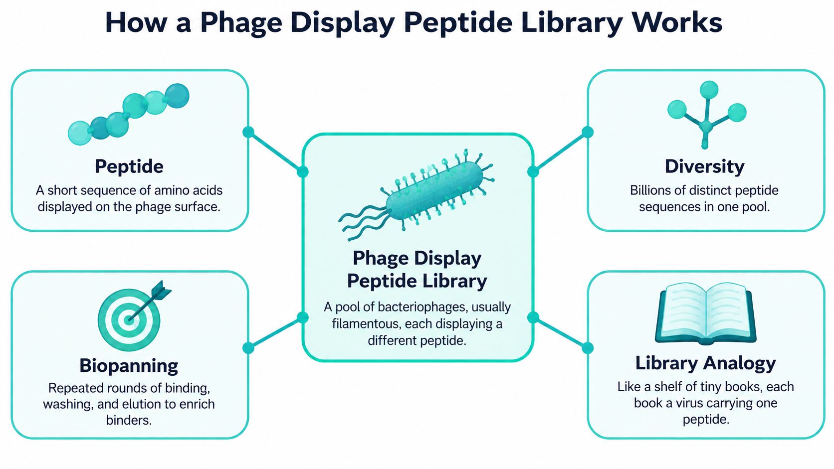
Researchers build the library, expose it to a target, wash away unbound particles, and recover those that remain. They then amplify the recovered phage in bacteria and use that population for another selection. This repeated enrichment process is called biopanning.
Peptides suit this approach because researchers can vary their amino acid sequences through DNA design and read the resulting sequences by sequencing the inserts. A mixed library lets them examine far more candidates together than would be practical to synthesize and test separately.
A recovered phage has survived the screening conditions. That alone doesn't establish why it survived.
Check your mental model before proceeding: the peptide is displayed outside, its gene travels inside, and washing removes particles that don't remain associated under the chosen conditions. The eventual task is to connect that survival back to genuine peptide–target binding.
Say your laboratory has an antibody that recognizes a protein, but you don't know which part it recognizes. You screen the antibody against a peptide library, sequence the retained inserts, and compare recurring peptide patterns with the protein's sequence and structure.
A matching sequence can suggest a linear epitope, the region recognized by the antibody. However, a selected peptide can also mimic a binding surface without matching the original protein's sequence. Such a mimotope is a clue about recognition, not a direct map of the native binding site.
An epitope-mapping workflow connects plate-based screening with sequence interpretation.

The readout must answer the intended question. A receptor-binding peptide isn't automatically a signaling ligand, and a disease-associated antibody response isn't automatically a diagnostic test.
Every application produces research candidates that require validation in the relevant biological or materials setting. Moving toward clinical use requires separate analytical, biological, and clinical evaluation beyond a discovery screen.
Common phage display peptide library formats
| Format | Display Site | Best Fit |
|---|---|---|
| Linear random 7-mer | pIII fusion, with low-valency display achievable | Broad binder discovery and epitope mapping |
| Linear random 12-mer | pIII fusion, with low-valency display achievable | Longer inserts and larger binding surfaces |
| Cyclic constrained CX7C | pIII fusion, with low-valency display achievable | Conformation-dependent binders |
| Short peptide on pVIII | Major coat protein, potentially high-density display | Weak interactions where avidity helps detection |
The coat protein choice affects how the target encounters each peptide. With pIII-based systems, researchers can use display configurations that reduce opportunities for simultaneous contacts. With pVIII, the phage surface can present peptides more densely, although the actual fusion occupancy depends on the construct and production system.
That distinction separates affinity, the strength of an individual interaction, from avidity, the combined retention produced by multiple contacts. Dense display can help detect weak interactions, but it can also make an unimpressive individual binder appear convincing. Neither the coat protein name nor a supplier's format description establishes the actual display valency in your preparation.
A shorter linear insert provides a compact recognition surface. A longer insert offers additional contact possibilities, but it also has more conformations available, which can complicate transfer into a useful synthetic peptide.
A cysteine-flanked design such as CX7C is intended to form a disulfide-constrained loop. That constraint can help present a preferred shape, provided the disulfide forms and remains intact under the assay conditions. It isn't interchangeable with a chemically cyclized peptide using a different linkage.
Plan the eventual solid-phase peptide synthesis while choosing the library. Terminal attachments, cyclization chemistry, and residues contributed by the display junction can all influence what you should synthesize.
Selection rule: Choose the format that can answer your binding question, not the format with the largest advertised library.
The theoretical sequence space isn't the same as the population present in your tube. Transformation, sequence bias, propagation, and sampling determine which variants are available. Specialized alternatives, including T7 and lambda systems, also exist, but their assembly and display constraints require different design choices.
Design the insert and its junctions. NNK degenerate codons can provide broad amino acid coverage while reducing stop-codon opportunities relative to unrestricted NNN randomization; N represents any DNA base, and K represents G or T. Check the reading frame, secretion signal, coat-protein junction, and host compatibility, because a well-randomized insert is useless if the fusion cannot be expressed.
Convert the oligonucleotide into a double-stranded insert using the assembly method matched to your vector. For restriction-based cloning, verify complete digestion and clean up the products, since residual uncut vector can produce colonies that look like successful library members but contain no insert.
Optimize ligation before scaling. Compare insert-to-vector conditions in a pilot, including a vector-only control, and choose conditions that favor correctly assembled constructs. Adding more insert or increasing reaction volume won't rescue damaged ends, poor DNA quality, or incompatible cloning chemistry.
The process overview connects insert design with phage production.
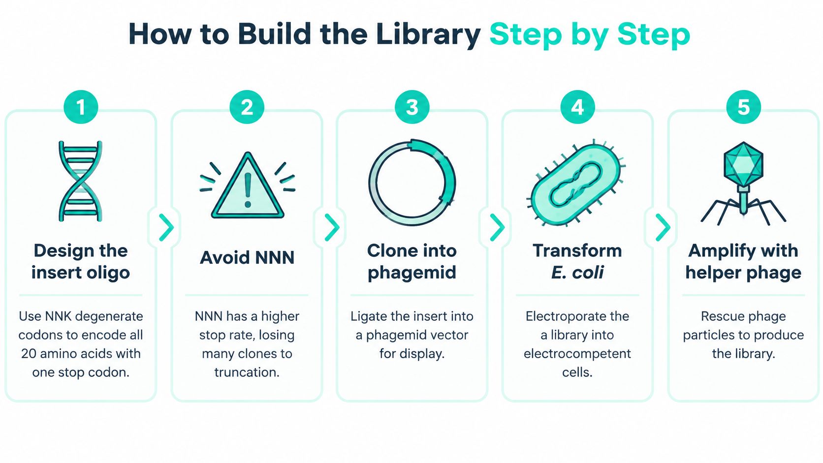
Electroporate the assembled DNA into a compatible, highly electrocompetent E. coli strain and recover the cells in SOC medium under validated host conditions. Salt carryover, excessive DNA loading, or compromised cells can sharply reduce the number of independent transformants, regardless of how ambitious the oligonucleotide design was.
Estimate the transformant population with dilution plating. Calculate colony-forming units from colony counts, dilution factors, plated volume, and total recovery volume, while using the vector-only control to assess background. Treat that estimate as a ceiling on independent library members, not proof that every transformant carries a unique, functional peptide.
For a helper-dependent phagemid system, rescue the library with a compatible helper phage and amplify under conditions that preserve representation. Recover particles using an established PEG and sodium chloride precipitation workflow or another validated purification method; poor rescue, uneven bacterial growth, and excessive amplification can distort the pool before selection begins.
Check sequences and preserve separate stocks. Sequence randomly selected clones to inspect insert presence, reading frame, stop codons, junctions, and unexpected sequence bias, then measure the infectious phage titer. Preserve the bacterial library in a suitable glycerol stock and store purified phage separately under validated conditions, since bacterial cryopreservation and phage storage aren't the same procedure.
A small clone sample can reveal obvious construction failures, but it can't establish full-library diversity. When representation matters, broader sequencing provides a more informative baseline for interpreting later enrichment.
Keep construction records with the stock identity. Without a trustworthy starting population, you can't tell whether a recurring sequence emerged through target selection or was already overrepresented.
Typical biopanning schedule by round
| Round | Elution | Expected Enrichment |
|---|---|---|
| Initial round | Acid elution if compatible with the system | Broad recovery, often with substantial background |
| Following round | Acid elution or a validated alternative | Candidate enrichment, with repeated sequences potentially appearing |
| Specificity-focused round | Competitive elution with free target when feasible | Preferential recovery of competitively displaced candidates |
| Additional rounds | Competitive elution, or stop when useful separation no longer improves | Possible convergence on a narrower set of motifs |
This is a planning framework, not a required progression. A change in elution method changes which binders you can recover, so don't interpret successive outputs as directly comparable without considering that change.
Immobilize the target on a plate or beads using chemistry that preserves the relevant binding surface. Passive adsorption is convenient, but it can alter a protein's conformation or hide an epitope against the support. Check target capture and activity before interpreting a quiet screen as evidence that no binders exist.
Block exposed surfaces with a compatible reagent such as BSA or milk, then introduce the library under conditions that preserve target stability. Include matched controls for the support, blocker, capture reagent, and any purification tag. A peptide that recognizes the target's affinity tag can otherwise look like a successful target binder.
Use counter-selection to remove unwanted binders before positive selection. Expose the pool to the blank support or relevant nuisance component, then transfer the unbound fraction to the target. Keep a record of what the negative selection removes, because an overly aggressive counter-selection can also discard useful candidates.
Washing removes unbound particles and challenges retained interactions. Change wash duration, detergent, salt, or other conditions deliberately rather than increasing every parameter together. Otherwise, you won't know which change removed your candidates.
Acid elution disrupts interactions broadly and requires prompt neutralization to protect phage infectivity. Competitive elution with soluble target can favor candidates displaced through the intended interaction, but it may miss binders that dissociate slowly or cannot access the soluble target configuration.
More recovered phage doesn't necessarily mean better binding. Recovery also reflects background retention, elution efficiency, infectivity, and bacterial amplification.
Measure infectious input and recovered output before amplification, and compare target recovery with the matched negative controls. An increasing output-to-input ratio supports enrichment only when the target-specific separation improves too.
Amplify recovered particles for the next round while limiting propagation bias. Sequence intermediate populations when possible, and stop when additional selection no longer improves the useful distinction between target and control. A dominant clone deserves investigation, not automatic celebration: it may be a strong binder, a background binder, or an efficient propagator.
Enrichment isn't proof that a free peptide binds. Begin by grouping sequences into exact matches and related motif families, while comparing them with the starting pool and negative-selection outputs.
An alignment helps you choose representatives rather than repeatedly testing nearly identical candidates.

Select candidates across distinct sequence families. Hydrophobic stretches, strong charge, or recurrence in negative controls warrant investigation, but no sequence feature alone proves nonspecific binding. Independent selections provide stronger support than repeated recovery after amplification from a shared ancestor.
Synthesize candidates with the intended termini and any required constraint. Confirm identity by mass spectrometry and assess purity chromatographically; understanding peptide separation techniques helps distinguish a sequence problem from an impure or heterogeneous preparation.
Test the target alongside relevant control proteins, blank surfaces, and tag-only controls. If detection requires biotin or a fluorophore, compare an unlabeled competitor where practical, because the label and attachment site can change behavior.
The accompanying video provides another visual reference for the display approach.
Use ELISA for initial comparison, then an appropriate method such as surface plasmon resonance or bio-layer interferometry to examine concentration-dependent binding. Estimate an equilibrium dissociation constant, KD, only when the assay design and binding model support it; immobilization artifacts and mass-transport effects can undermine the fit.
A free peptide may lose the favorable presentation supplied by the phage, including neighboring residues, tethering, or multivalent contacts. Check solubility and chemical integrity before redesigning it, then test rational alternatives such as a junction-matched extension or a correctly constrained version.
Advance candidates only when the synthetic molecule shows reproducible, target-specific binding. A modified peptide that succeeds is a newly validated construct, not retrospective proof that the original free sequence worked.
Low diversity limits the experiment before target selection begins. Check construction quality before spending effort adjusting washes.
Symptoms, causes and fixes
| Symptom | Likely Cause | Fix |
|---|---|---|
| Few transformants, diversity far below design | Weak electrocompetent cells or poor ligation | Check cell competence and DNA quality; optimize insert-to-vector conditions |
| Output titer does not rise across rounds | Poor target presentation, harsh washes, or inefficient recovery | Confirm target activity and capture; test gentler washing and recovery |
| A clone dominates early | Strong selection or a propagation jackpot | Compare target and control binding; inspect intermediate sequences and shorten amplification |
| Hits bind plastic or the blocking agent | Inadequate counter-selection | Add negative selection against the support and blocker |
| Hits fail as free peptides | Avidity, scaffold dependence, or peptide quality problems | Verify material quality; test justified constrained or extended versions |
| Many clones carry no insert | Empty-vector background | Improve digestion or assembly; dephosphorylate vector when compatible with the cloning strategy |
Poor transformant recovery points toward the library-building workflow. Adequate construction quality with poor target-specific recovery shifts attention toward target presentation, assay conditions, and elution.
A propagation jackpot is especially misleading because it can resemble successful selection. Comparing sequence abundance before and after amplification helps reveal whether bacterial growth, rather than target contact, drove the takeover.
Record transformant estimates, infectious titers, target preparation, wash conditions, elution conditions, and sequence identities together. These records let you identify the stage where the experiment diverged instead of repeating the entire workflow blindly.
A useful screen starts with the intended final reagent. Decide whether you need a soluble probe, an immobilized capture peptide, or a constrained inhibitor candidate before committing to a display format.
Define the target state. Specify the relevant conformation, surface, cofactors, and presentation method. Decide which controls can distinguish recognition of the target from recognition of its support or tag.
Choose the library architecture for that question. Favor a compact linear design for short recognition features, a constrained design when shape matters, and dense display only when you can separately evaluate its contribution to detection.
Write the selection logic. Establish counter-selection, wash adjustments, recovery methods, and stopping criteria before screening. Change conditions to answer a question, not just to make each round harsher.
Track sequences through selection and amplification. Choose synthesis candidates that represent distinct families and plausible binding mechanisms rather than only the most abundant output.
Budget for chemical and binding validation. Use HPLC and mass spectrometry analysis to establish what material you're testing, then evaluate specificity and affinity with suitable controls.
Preserve the experimental record and the relevant stocks. Another researcher should be able to reconstruct both how you obtained a candidate and why you considered it worth advancing.
We design our research peptides for scientific study in laboratories worldwide. All our peptides carry a research-use-only designation, which is separate from demonstrating suitability for any particular binding experiment.
For a practical pilot this week, use a well-characterized target with a known binder and matched negative controls. Confirm that target presentation, recovery, and detection behave as intended before committing your discovery library to an unfamiliar system.
Celonyx Labs offers research peptides for laboratory investigation, where material identity and experimental controls matter as much as candidate selection. Visit Celonyx Labs to explore the research peptide catalog and supporting information while planning your peptide experiments.
The advice you'll hear most often about buying BPC-157 for the bench is short: demand

Can cagrilintide be combined with GLP-1 peptides? Yes, in controlled research. Understand mechanisms, study controls,

Can Selank be combined with nootropic peptides? Yes, in controlled laboratory studies. Learn the controls,
FOR RESEARCH USE ONLY. Products sold by Celonyx Labs are intended exclusively for laboratory and scientific research. They are not intended for human or veterinary use, consumption, diagnosis, treatment, cure, or prevention of any disease. By purchasing from this site, you acknowledge that you understand the intended research purpose of these materials and agree to comply with all applicable laws and regulations.