

BPC 157 500mcg Explained: What the Evidence Shows
500 mcg equals 0.5 mg, a labeled research quantity, and no published human study establishes
You're standing at the bench with a peptide vial, a vendor purity claim, and a project that can't afford a wrong start. The sample looks clean enough, the paperwork looks polished, and yet you still need to know whether the material is really what it says it is, whether the main peak is actually your peptide, and whether the trace signals are real impurities or just contamination from the way the run was handled. That's where HPLC MS analysis earns its place in peptide research, because it turns a catalog claim into evidence you can defend.
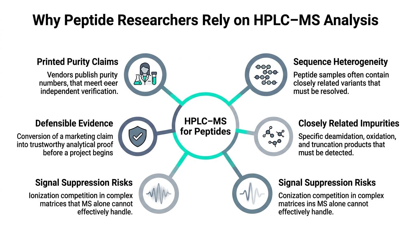
A peptide researcher often starts with a simple question. The vial says one thing, the certificate says another, and the experiment waits on the answer. A UV trace by itself can suggest purity, but it can't tell you whether a peak is really the expected peptide, a closely related truncation product, or an impurity that happens to absorb in the same place.
That's why HPLC‑MS analysis is so useful in this workflow. The chromatographic part separates components that would otherwise overlap, and the mass spectrometric part identifies what each component is by mass-to-charge ratio. The combination matters because peptide samples rarely behave like a single neat compound, they usually contain sequence variants, oxidation products, deamidation products, residual synthesis byproducts, and matrix-related noise that can obscure the answer if you rely on only one technique.
A vendor's purity statement is not the same thing as independent evidence. The key question is whether the major signal belongs to the expected peptide and whether the minor signals make chemical sense. HPLC‑MS is the practical bridge between those two needs, because it gives you both a separation trace and a molecular identity check in one workflow, which is especially valuable for impurity profiling and identity confirmation in the same run, as described in the chromatographic separation and mass-selective detection model from Clinisciences' overview of HPLC principles (Clinisciences).
For peptide work, that matters before any biologic assay, binding study, or formulation trial begins. If the starting material is wrong, the downstream data can still look polished and be completely misleading.
Practical rule: if the peptide hasn't been checked by a method that separates components and reads their masses, the printed purity number is still just a claim.
A good way to think about it is this, HPLC tells you what came off the column and when, while MS tells you what that thing weighs. For a peptide scientist, that's the difference between trusting a label and verifying the chemistry.
The easiest way to understand the instrument is to treat it as separation, then weighing. HPLC first spreads the peptide mixture out across time. MS then examines each slice of that flow and asks what molecular species are present.
In peptide analysis, the column is usually doing reversed-phase work, which means the peptide's interaction with the stationary phase influences when it elutes. More hydrophobic peptides often stay longer, while more polar ones may elute earlier. The important point isn't the exact chemistry of every stationary phase, it's that the column prevents everything from arriving at the detector as one blur.
Once components are separated, the ion source turns molecules in the liquid stream into gas-phase ions. In peptide work that usually means electrospray ionization, which is useful because peptides can carry multiple charges. That charge state is what lets the mass spectrometer measure them by m/z, not just by neutral mass.
A mass analyzer then sorts those ions. In a full-scan run, you get a broad view of what ions are present. In MS/MS, the instrument selects one ion, fragments it, and reads the pieces. That second step is often what gives you stronger identity confidence, because fragment patterns can support the sequence you expected.
The distinction matters because HPLC alone can show a single sharp peak that still belongs to the wrong molecule, and MS alone can detect the right mass in a crowded background without telling you whether it co-eluted cleanly. Hyphenating the two removes that blind spot.
A chromatogram without mass data tells you about retention behavior and apparent purity. A mass spectrum without chromatographic separation tells you about ion composition at a moment in time. Together, they answer both questions at once, which is why the field treats HPLC-MS as one analytical system rather than two instruments bolted together.
The logic is simple. The column handles complexity in time, and the mass spectrometer handles complexity in identity. That's the combination peptide researchers need when a single vial must stand in for a whole project.
The stronger the sample complexity, the more valuable it is to separate before you identify.
The bench layout is usually familiar once you've seen it once. A binary pump drives the gradient, an autosampler injects the sample, the column oven steadies retention, the ion source makes the ions, and the mass analyzer sorts them before the detector records the result. For peptide work, the parts that matter most are the ones that influence separation quality and ion formation, not just the headline instrument name on the brochure.
Column chemistry shapes what you can separate. C18 is the usual starting point, but C4 and diphenyl columns can be useful when a peptide's hydrophobic behavior or aromatic interactions need a different selectivity. Gradient steepness matters too, because a gradient that is too aggressive can compress closely related peptide species into a crowded region, while one that is too shallow can stretch the run longer than necessary without adding useful resolution.
Ion-source conditions also matter more than many newcomers expect. If the source is too harsh, you can lose signal or create unstable spectra. If it's too soft or contaminated, ion formation becomes inconsistent. That's why peptide labs often spend more time tuning source behavior than they first expect.
A single quadrupole can be useful for straightforward checks, but it gives limited selectivity. A triple quadrupole is especially useful when targeted monitoring is the goal, because it can isolate a precursor ion and track fragments in a controlled way. High-resolution platforms such as time-of-flight and Orbitrap systems are often chosen when accurate mass and cleaner identification are central to the workflow.
The operating mode should match the question. Full scan asks what ions are present. Selected ion monitoring asks whether a specific ion is there. MS/MS asks whether the fragments support the expected structure. That progression is more useful than memorizing a feature list, because the underlying decision is always analytical.
Rule of thumb: choose the mode that matches the decision you need to make, not the most impressive setting available.
If the purpose is identity confirmation plus impurity detection, the method often leans toward separation quality first, then a mass mode that can support confident interpretation of both the main component and minor species.
The video below is a useful visual companion for how the instrument chain fits together.
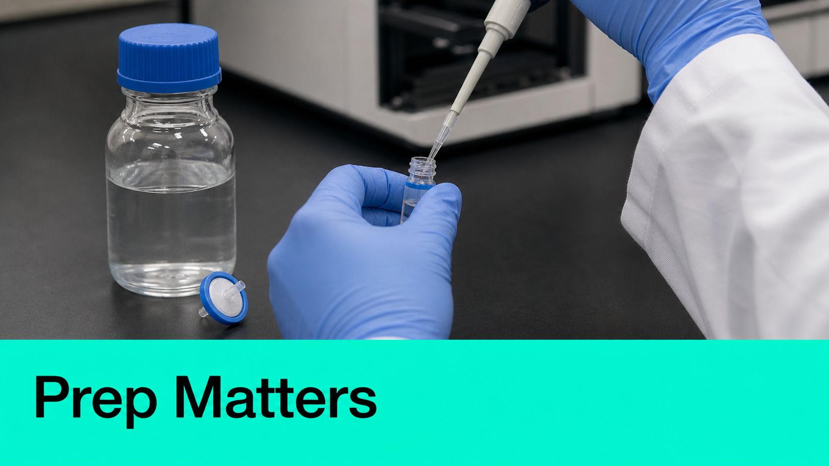
A peptide can fail before it ever reaches the column. Poor dissolution, adsorption to plastic, incomplete reconstitution, or an incompatible buffer can all distort the result before the first signal appears. That's why sample prep deserves the same seriousness as the instrument method itself.
Hydrophobic peptides often need stronger organic content to dissolve, while more hydrophilic or basic sequences may behave better in aqueous mixtures with a compatible modifier. The peptide's sequence and charge properties should guide that choice, not whatever solvent was on the bench first. A sample that looks dissolved can still contain invisible aggregation or surface adsorption that affects injection behavior.
Additives change the story quickly. Formic acid, trifluoroacetic acid, and ammonium buffers can all influence peak shape and ionization, sometimes in helpful ways and sometimes in ways that suppress signal. The key is matching the additive to the analytical goal. A peptide that needs maximum MS response may not tolerate the same solvent conditions as one being judged mainly by chromatographic shape.
Filtration still matters, even for solutions that look clean. Tiny particulates can damage the column, distort peaks, or create background issues that look like chemistry problems when the real issue is bench handling. For practical reconstitution choices, a focused lab guide such as the one on peptide reconstitution workflows can help connect theory to handling.
A sample prep step that seems routine can decide whether the run is interpretable or misleading. For peptide analysts, that's not a minor detail. It's the difference between a result you can trust and a result you'll end up redoing.
The chromatogram is the first place overreading and underreading often occur simultaneously. Use the step-by-step HPLC chromatogram guide to check retention time, integration boundaries, peak shape, and baseline behavior before assigning purity or identity. A peak is visible and immediately associated with purity. In reality, the peak is a record of retention behavior, and its shape provides significant insight into how the peptide moved through the system.
Retention time tells you when the component eluted. Peak width tells you how dispersed it was. Tailing often suggests interaction problems, overloading, or surface effects, while a noisy baseline hints at system issues, contamination, or unstable mobile phase behavior. These are not abstract diagnostic terms, they are the first clues that the column, sample, or solvent system needs attention.
The mass spectrum adds the next layer. Peptides often appear as a charge-state envelope, meaning several charged forms of the same molecule may be visible at once. That's normal in electrospray. The spectrum is therefore less like a simple weighing scale and more like a pattern you have to interpret before you can assign a neutral mass.
A base-peak chromatogram shows the most intense signal at each point in time, which is useful for an overview but not always for specificity. An extracted-ion chromatogram focuses on one m/z value or a narrow mass window, which helps isolate the peptide of interest from background species.
Deconvolution takes the multiple charged ions and translates them into a single neutral mass. That step is what lets a peptide researcher compare the observed species against the expected sequence mass. When the main peak's mass lines up and the minor peaks fit a plausible impurity pattern, the data becomes much easier to defend.
Practical interpretation: a clean peak with the wrong mass is not a clean result.
If the main chromatographic peak produces the expected neutral mass, that supports identity. If a second peak sits nearby and deconvolves to a slightly different mass, that could indicate a modification such as oxidation or deamidation, or it could be a system artifact. The difference comes from whether the mass shift and the retention behavior make chemical sense together.
That's why chromatogram and spectrum need to be read as a pair. One tells you when, the other tells you what. In peptide work, neither one is enough by itself.
A strong signal feels convincing. It isn't always quantitative. That's the trap many peptide researchers run into when they treat presence and abundance as the same question.
The open literature increasingly points to this distinction in LC-MS workflows. A method can be excellent at confirming that a peptide is present at the expected mass, yet still be weak for estimating how much of it is really there, especially in complex biological or peptide-adjacent samples. As noted in recent LC-MS workflow guidance, matrix effects, ion suppression, extraction variability, and run-to-run drift are often the actual bottlenecks, not detection alone (PMC review on LC-MS workflow robustness).
That's why internal standards matter. They give the method a reference point so signal changes can be judged against something stable rather than against the raw instrument response alone. In targeted work, MS/MS acquisition and ratio-based normalization are practical ways to make the number more defensible when the question really is quantitation rather than mere identification.
A purity claim based on one UV trace without mass confirmation is weaker than it looks. The UV peak may be real, but without mass data you don't know whether the signal includes co-eluting impurities or whether the apparent abundance is distorted by suppression and extraction differences. For complex samples, a number deserves trust only when the method can justify it.
Use this decision frame:
For peptide researchers who want a practical impurity-focused perspective, this guide to peptide impurity profiling methods is a useful complement to the analytical logic here.
The key is not to force every HPLC-MS run into a quantitation claim it can't support. A clear identification can be enough for one project stage, but if the number is going to drive the next decision, the method has to earn that number.
Most “bad peak” problems aren't dramatic instrument failures. They're small habits that accumulate until the run gets noisy, weak, or misleading. A recent review of LC-MS performance emphasizes fresh mobile phases, pH control with appropriate modifiers, column conditioning and regeneration, metal deactivation, and sample-prep discipline, while vendor guidance also stresses the use of MS-grade solvents and ultrapure additives and warns that dead volume, solvent contamination, and microbial growth in aqueous mobile phases can materially reduce sensitivity (Analytical Chemistry Research review).
A solvent bottle cap left open too long can invite airborne contamination. A mobile phase that sits until microbial growth starts changing the system can degrade sensitivity. Extra dead volume in tubing and fittings can smear peaks and make the chromatogram harder to read. Metal surfaces in the flow path can also matter, especially when the peptide or modifier chemistry makes the system more reactive than expected.
These aren't glamorous troubleshooting topics, but they're often the first things to check when the signal changes without a method change. If the column and source are blamed before the bottles, the tubing, and the cleaning cadence, the problem can linger for days.
If sensitivity drops and the method hasn't changed, inspect the bench before you suspect the analyzer.
The useful mindset here is simple. Data quality is cumulative. Every small handling choice either preserves the chemistry or erodes it a little more, and peptide analysis is sensitive enough to show the difference.
A credible third-party report should let another scientist understand what was done and why the result is believable. If it doesn't, the purity number is unfinished.
At minimum, the report should identify the chromatographic method, the column, the gradient, the ionization mode, and the mass range or transitions monitored. It should also show how peaks were integrated, what thresholds were used, and how the mass spectrum confirms that the main peak matches the expected peptide mass. Without those details, the result is hard to audit and impossible to compare cleanly against another lab's work.
That matters when evaluating a Certificate of Analysis. A figure that says “99% purity” on its own is not the same as a trace, a spectrum, and conditions that let you judge how that number was obtained. Third-party testing should behave like an audit, not a branding exercise. A vendor page such as the one describing third-party tested peptides is useful to the extent that it points a reader toward that standard, but the report itself is what proves the claim.
Look for a match between the claimed peptide and the observed mass. Check whether the chromatogram is shown, not just summarized. Look for whether the method was targeted enough to separate the main species from obvious nearby peaks. If the document omits conditions, the reader can't judge how hard the method worked to earn the purity call.
A good report answers these questions plainly:
That is what makes a purity report useful to a peptide researcher. It doesn't just state a number, it explains the path from sample to answer.
If you want research peptides backed by clear quality information and independent third-party testing, visit Celonyx Labs and review the catalog with the same questions you'd ask of any analytical report. A good peptide supplier should make verification easier, not harder, and Celonyx Labs is built around that expectation for laboratory buyers who need dependable starting material.
500 mcg equals 0.5 mg, a labeled research quantity, and no published human study establishes

Choose the best GHK-Cu peptide format for laboratory skin and wound-healing research. Compare documentation, assay

Choose lot-verified melanotan 1 peptide for pigmentation research. Compare material specifications, purity evidence, and documentation
FOR RESEARCH USE ONLY. Products sold by Celonyx Labs are intended exclusively for laboratory and scientific research. They are not intended for human or veterinary use, consumption, diagnosis, treatment, cure, or prevention of any disease. By purchasing from this site, you acknowledge that you understand the intended research purpose of these materials and agree to comply with all applicable laws and regulations.